![]()
![]()
![]()
![]()
The Optics Laboratory
Group of
Hans Hallen, North Carolina State University Physics Department![]()
Related Techniques
• Light in/out of Uncoated Probes
• Light Detection at the Probe Tip
• Apertureless NSOM
• Photon Tunneling Microscopy (with a Solid Immersion Lens)
• Confocal Microscopy, Etc.
Light in/out of Uncoated Probes
• G. Krausch, et al. Opt. Comm. 119 (3-4) 283-8 (1995).
• W.A. Atia et al. CLEO '95, 213-14 (1995).
• A. Jalocha, et al. Ultramicroscopy 61 (1-4) 221-6 (1996).
• G. Kaupp, A Herrman, M. Haak, J. Vac. Sci. Technol. B15 (4)
1521-6 (1997).
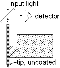
• An 'easy' way to get ~
• Get large signals since have no waveguide cut-off.
• Like NSOM but light is both input and collected through the fiber, which is uncoated here.

• Will have a large amount of 'un-used light' on the sample compared to NSOM (can bleach fluorescent molecules quickly as confocal can).
Light Detection at the Probe Tip
• D. Busath, R. Davis and C.C. Williams, SPIE 1855, 75-80 (1993).
• R. Davis, C.C. Williams and P. Neuzil, APL 66 (18) 2309-11
(1995); and Opt. Lett. 21 (7) 447-9 (1996).
• G Kolb et al, Ultramicroscopy 57, 208-11 (1995).
• H. Yamada et al, J. Vac. Sci. Technol. B14 (2) 812-15 (1996).
• R.G. Davis, C.C. Williams and P. Neuzil, APL 69 (9) 1179-81 (1996).
• Typically a semiconductor photodiode is fabricated at the tip of a silicon AFM probe on a cantilever.
• Have a high conversion efficiency compared to the fiber probes.
• Complement NSOM since the direct absorption of power in the near field is different.
• Still must be careful about topographic coupling in resolution measurements.
Apertureless NSOM
• F. Zenhausern, M.P. O'Boyle and H.K. Wickramasinghe, APL 65 (13) 1623-5 (1994).
• F. Zenhausern, Y. Martin and H.K. Wickramasinghe, Science 269 (5227) 1083-5 (1995).
• S. Kawata, Y. Inouye, Ultramicroscopy 57, 313-17 (1995).
• R. Bachelot, A. Lahrech, P. Gleyzes, and A.C. Boccara, SPIE 2782, 570-81 (1996); Ultramicroscopy 57, 318-22 (1995).
• Instead of a clear aperture, use a tip to block/ interact with light (from far field) in a local position
• Oscillate the tip vertically and lock-in to select the interacted light (there is a large background illumination).
• Most resolution claims are from regions with topography, but single molecule spot sizes are ~10 nm -- as good as the best aperture NSOM.
Can also be done scattering from particles rather than a tip.
• T.J. Silva and S. Schultz, RSI 67 (3) pt 1, 715-25 (1996).
• and other work by the same authors.
• The particles are scattered and imaged or it can be moved with a probe or optical tweezers.
• They have concentrated on magnetic measurements, but the technique is more general.
Photon Tunneling Microscopy with a Solid Immersion Lens
• John M. Guerra, Mohan Srinivasarao and Richard Stein, Science 262, 1395-1400 (1993).
• John M. Guerra, MRS Proc. 332, 449-460 (1994).
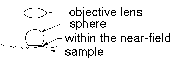
• Uses the fact that the wavelength hence diffraction broadening of the focus are reduced in a material with index of refraction greater than one.
• This solid immersion lens surface must be within the near-field of the object for the effect to be applicable.

• The shape of the sphere must be nearly perfect to reduce aberrations so that improved resolutions are achieved.
• Such a lens can be flown over a surface as a hard-disk head is: B.D. Terris, H.J. Mamin, and D. Rugar, APL 68 (2) 141 (1996).
Confocal Microscopy, Etc.
• Instead of an aperture in the sample plane as NSOM, the aperture is scanned in the back focal plane.
• Lateral resolution is far-field diffraction limited.
• The depth resolution is set by the depth of focus of illumination/ collection optics and aperture size, and can be very fine.
Combined with interference
• M. Vaez-Iravani et al, Ultramicrosc. 61, 105 (1995).
• Can achieve very high lateral resolution.
Combined with two-photon microscopy (4-
• S.W. Hell, M. Schrader, and H.T.M. van der Voort, J. Microsc. 187 (1) 1-7 (1997).
• Gains additional resolution since the 2-photon effect further limits the important focal spot.
• Still diffraction limited.
• Warning: sometimes maximum entropy (all- poles) deconvolution techniques are used to enhance resolution. These algorithms assume that the image is made up of sharp spikes or lines, so return such sharp features even if the sample actually has smoother features. Their width is a measure of the algorithm not the instrument resolution.
![]()
Last updated on September 27, 2000