![]()
![]()
The Optics Laboratory
Group of
Hans Hallen, North Carolina State University Physics Department![]()
![]()
![]()
The Optics Laboratory
Group of
Hans Hallen, North Carolina State University Physics Department![]()
Near-field scanning optical microscopy (NSOM, also known as SNOM, NFOM) elucidates to the interaction of light with a sample close to a metal aperture, which constrains the lateral extent of the light. The aperture is held in place in a manner similar to those used for other scanning proximal probe microscopes. Advantages of the near-field interactions are:
These properties can be utilized in a variety of experiments.
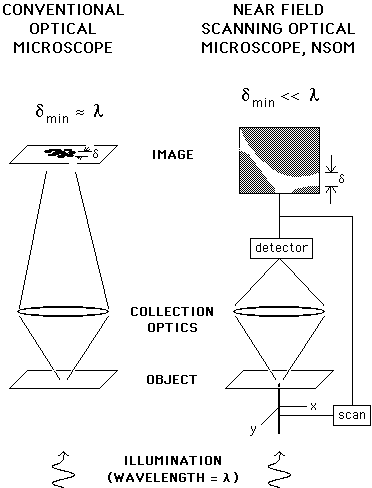
A generic NSOM looks roughly like this:
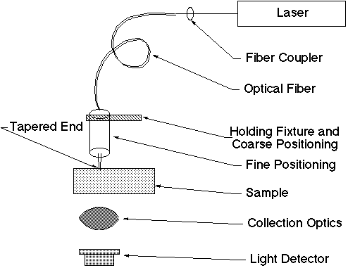
The microscope itself is in the middle, sometimes including the detectors and sometimes not. A single mode fiber, and hence the laser light source (think étendue), are external.
Some History:
• Conception of an instrument with most modern features. - E.H. Synge, "A suggested method for extending the microscopic resolution into the ultramicroscopic region," Phil. Mag. 6, 356 (1928). - E.H. Synge, "An application of piezoelectricity to microscopy," Phil. Mag. 13, 297 (1932).
• Experiment in the microwaves. - E.A. Ash and G Nichols, "Super-resolution aperture scanning microscope," Nature 237, 510 (1972).
• Experiments in the visible. - D.W. Pohl, W. Denk, and M. Lanz, APL 44, 651-3 (1984). - A. Lewis, M. Isaacson, A. Harootunian, and A. Murray, Ultramicroscopy 13, 227 (1984); Biophysics Journal 41, 405a (1983).
E. Betzig, et al. Biophys. J. 49, 269 (1986).
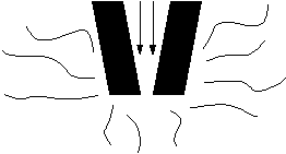
Before we understand near-field resolution, consider the limits of far-field optics, due to far-field diffraction:
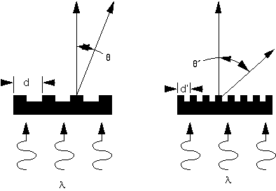
If the lens does not collect the first order diffracted light described by
l
= d sinq,then the surface features are not seen. A Fourier transform analysis connects a real-world image with the simple grating — the closely-spaced features are not seen. The near-field instrument uses geometry to confine the light:
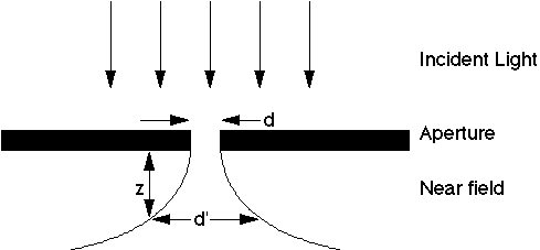
Here,
d <<
l,and the light lines represent the spreading of the field. The integral of the field along the horizontal between the lines is the same at any vertical height. Of note is that the wavelength doesn’t effect the resolution (except as the optical properties of the aperture material vary). It does, however, enter into the calculation of how much light passes through the aperture — more on that later.
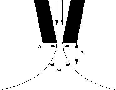
For NSOM we need to get close to the sample, so a point is preferred to a flat aparture, giving an NSOM coated, tapered, optical fiber probe:
• For high spatial resolution, the probe must be close to the sample.
• Some (preferably independent) feedback mechanism must be used to hold it there.
The experimentalist's view:
• Operating Modes
• For high spatial resolution, the tip must be close to the sample.
• How does it compare to far-field microscopy?
• Why do we not get contrast from subsurface structures?
• How does the tip effect the measurement? - optically active states in the sample can be modified. - evanescent fields near the tip can couple into the sample effectively giving a (distance dependent) increase in the number of photons.
• Spatial resolution
• Contrast mechanisms
• An instrument example
Modes of operation
• Several modes offer flexibility in amount of light incident on the sample, microscope geometry, sample characteristics (opaque, transparent, etc.).
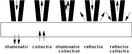
• Illumination by the tip is probably the easiest to interpret, and gives the most signal. It requires a transparent sample, though, so can’t always be used.
• Reflection modes give less light, and are more dependent on the details of the probe tip, but allow one to study opaque samples.
• The illumination/collection mode provides a complement to the reflection modes, but the signal contains a large background from the light reflected without ever making it out of the probe tip. (This is removed by oscillating the tip vertically and measuring the signal at that frequency.)
Comparison to far-field imaging
Note that the conventional microscope gets the entire image at once, while that from the NSOM is built up point-by-point. A closer comparison (not shown) is between confocal microscopy and NSOM. In confocal, an aperture is scanned in the back image plane. In NSOM, it is smaller and scanned in the sample plane.
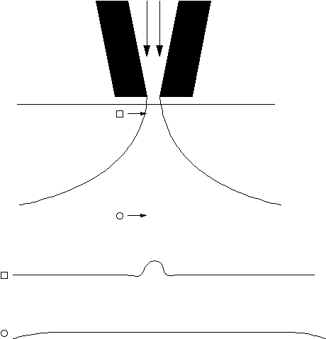
Here is a simple way to understand the surface sensitivity, just look at the amount of light present at each point:
Contrast, Surface vs. Subsurface:
Probe - sample interactions.
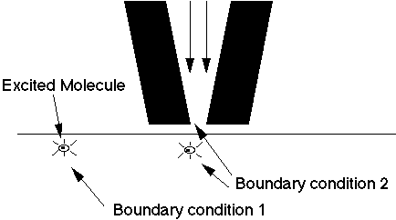
• Does the close proximity of the metal change the lifetime of the excited molecule?
YES:W.P. Ambrose et al, Science 265 (5170) 364-7 (1994).
R. Bian, R.C. Dunn, X.S. Xie, and P.T. Leung, Phys. Rev. Lett 75 (26) 4772-5 (1995).
Heating of the sample
• Recall that the limit to the power one can input to the fiber is determined by the melting of the aluminum coating (most light is reflected back).
• Energy is deposited during this reflection, so the probe tip gets very hot.
• The close proximity to the sample invites heat transfer (the heat diffuses through the layer of adsorbed material the tip rides in -- linear in the probe temperature and tip-sample separation). • Since the heated volume is very small (in its depth as well as width), thermal expansion effects in the sample are expected to be small. (the tip expands, however - La Rosa & Hallen)
• Reflection coefficients may depend on the temperature, especially in semiconductors.
• Some samples are changed by too much heat. P.J. Moyer, K. Walzer and M. Hietschold, APL 67 (15) 2129-31 (1995) found changes in liquid crystals.
• Biological samples (even cells) seem to be O.K. E. Betzig et al, Bioimaging 1 (3) 129-35 (1993) and many others since.
Evanescent photons from the tip
- can couple into the sample if it is close enough.
- can couple to propagating modes with large values of parallel momentum.
- results in surface enhancement (C. L. Jahncke and H. D. Hallen, 9th annual meeting of IEEE Lasers and Electro-Optics Society (LEOS) 96 conference proceedings volume 1, pp. 176-177.)
Spectroscopic Implications
• Near-field selection rules may differ from their far-field counterparts. above Jahncke and Hallen LEOS paper above, etc.
• Indirect transitions may be observable. Bob Grober, NFO-2 Proceedings, Ultramicroscopy 57 (1) (1995)
A nice experiment: B. Hecht, H. Heinzelmann, D.W. Pohl, Ultramicroscopy 57, 228-34 (1995).
• Light is coupled from a tapered fiber probe into a half-cylinder.
• Light intensity is measured as a function of propagation angle in the glass.
• Light is observed beyond the angle predicted for far-field illumination (from evanescent)
• The intensity drops as the tip is retracted.
Probe far from the surface:
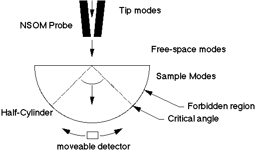
• Tip modes couple to free space modes, which couple to the sample.
• The detector gets no signal beyond the critical angle
Probe in the near-field:
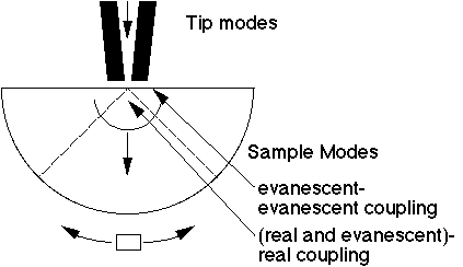
• Tip modes (including those usually evanescent) couple directly to the sample modes (including those usually evanescent).
• The detector receives light even beyond the critical angle (intensity exponential with tip to sample separation).
• The signal for angles smaller than critical is much less dependent on tip-sample separation.
Spatial resolution and contrast.
• These are coupled in NSOM
• Probe - sample separation is also an issue.
What does spatial resolution mean?
• We sample to get an image.
• Thus we obtain a Nyquist limit to the resolution - We assume here that the data is taken with small enough steps that other limitations apply.
Resolution:
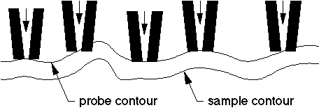
• Topographic induced contrast in an optical image often gives the appearance of higher resolution than can be achieved with the same probe on a flat surface. 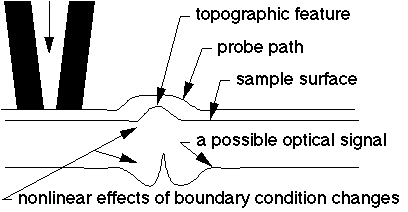
• Since the dependence is nonlinear, it cannot be modeled as a simple convolution.
• Resolution is tough to measure experimentally.
• Don't take all claims literally.
• Correlate your optical and topographic images.
• Test that your optical images depend upon intensity, distance and wavelength as they expected.
• Don't crash.
• E. Betzig and J. Trautman, Science 257, 189-95 (1992).
• S.I. Bozhevolnyi, JOSA B14 (9), 2254-9 (1997).
• B. Hecht et al, JAP 81, 2492-8 (1997).
Contrast Mechanisms
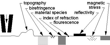
• Several contrast mechanisms exist.
• Most have been demonstrated.
• Experimental techniques - intensity - polarization - wavelength dependence - source wavelength - time - force feedback
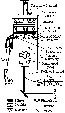
A microscope with some of its novel features.
C.L. Jahncke and H.D. Hallen, "A versatile, stable scanning
proximal probe microscope," Review of Scientific Instruments,
68 (4), 1759 (1997).
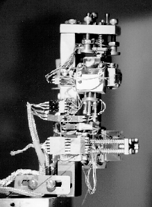
A photograph of the instrument.
![]()
North Carolina State University | Physics | Optics Home
Last updated on September 27, 2000