![]()
![]()
The Optics Laboratory
Group of
Hans Hallen, North Carolina State University Physics Department![]()
![]()
![]()
The Optics Laboratory
Group of
Hans Hallen, North Carolina State University Physics Department![]()
Applications
• Single Molecule Detection
• Raman Scattering
• Polarization
• Magnetic Imaging
• Data Storage
• Biological Imaging
• Quantum Dots, Quantum Lines
• Lithography
• Photonics Device Characterization
• Semiconductors/ Defect Characterization
Single Molecule Detection
• Some words about words: - The molecule is detected (signal).
- Molecules are only resolved if they are separated by a distance ~aperture size.
- The field profile of the tip is imaged.
- Photobleaching gives rise to quantized downward jumps in the signal.
- Calculations of signal levels.
- Comparison of number of molecules detected with density (varied) applied to sample.
- Modeling of the tip field profile interacting with a dipole.
- One can even measure the molecular orientation this way.
• Why NSOM? - Reduced background signal compared to far- field techniques.
- Longer time until photobleaching -- only one molecule is illuminated.
- Other techniques have also detected single molecs.
• Who in the early days? Many at NFO-2
- AT&T E. Betzig and R.J. Chichester, Science 262 (5138) 1422-8 (1993). J.K. Trautman et al. Nature 369, 40-2 (1994).
- Pacific Northwest Laboratory X.S. Xie and R.C. Dunn, Science 265, 361-4 (1994).
- LANL W. P. Ambrose et al. PRL 72, 160 (1994). W. P. Ambrose et al. Science 265, 364-7 (1994).
- ETH Zürich W.E. Moerner et al. PRL 73 (20) 2764-7 (1994).
• Recent work:
- In polymer films, not on the surface. M.A. Bopp, G. Tarrach, M.A. Lieb, and A.J. Meixner, J. Vac. Sci. Technol. A 15 (3) pt 2, 1423-6 (1997).
- On a resonant sphere. D.J. Norris, M. Kuwata-Gonokami, W.E. Moerner, APL 71 (3) 297-9 (1997).
- Theory Chang Railing et al. JAP 81 (8) 3369-76 (1997).
Raman Scattering
• Input light of one color (color not crucial).
• Energy analyze out-coming light.
• Phonon scattering can induce small changes in the color.
• The Raman spectra thus reflects the vibration modes of the molecule or crystal.
• It can be used
- as a fingerprint for species identification.
- to measure the local environment.
- to measure stress (shifts in phonon frequencies).
• Weak.
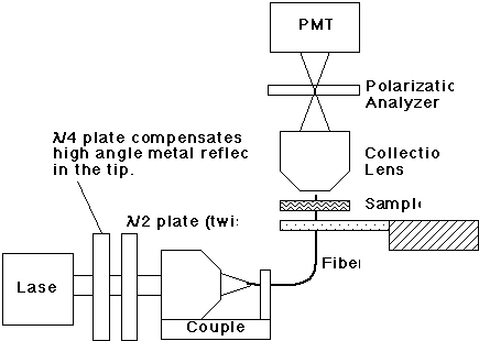
The experimental method:
• C.L. Jahncke, M. A. Paesler, and H.D. Hallen, "Raman imaging with near-field scanning optical microscopy," Appl. Phys. Lett. 67, (17), 2483-2485 (1995).
• H.D. Hallen, A. LaRosa, and C.L. Jahncke, "Near-field scanning optical microscopy and spectroscopy for semiconductor characterization," Physica Status Solidi (a) 152, 257-268 (1995).
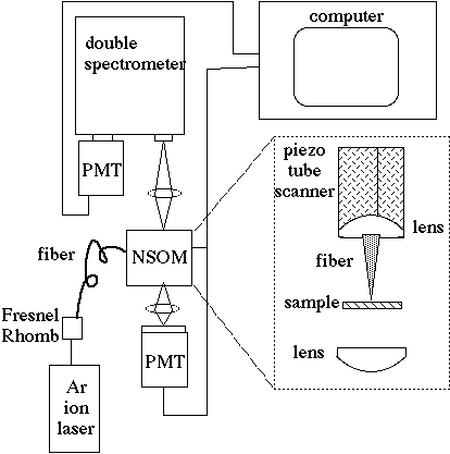
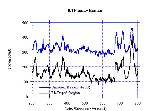
• Sample is KTP with Rb-doped regions.
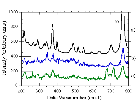
Raman spectra on a KTP sample:
• On and Off a Rb-doped region.
• C.L. Jahncke, H.D. Hallen, and M. A. Paesler, "Nano-Raman spectroscopy and imaging with the near-field scanning optical microscope," An invited contribution to the J. Raman Spectr. 27, 579-586 (1996).
• C.L. Jahncke, M. A. Paesler, and H.D. Hallen, APL 67, (17), 2483-2485 (1995).
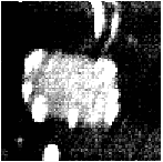
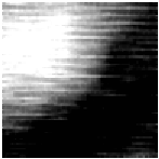
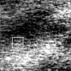
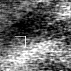
10 mm sq Topography 4 mm square NSOM transmission 4 mm square NSOM-Raman
Comparison of NSOM-Raman,
m-Raman, and Raman through a probe in the far-field• C. L. Jahncke and H. D. Hallen."Near-field Raman Spectra: surface enhancement, z-polarization, fiber Raman background, and Rayleigh scattering", 9th annual meeting of IEEE Lasers and Electro-Optics Society (LEOS) 96 conf. proc. 1, pp. 176-177.
Longitudinally Polarized Light in NSOM-Raman Spectroscopy
(a change in selection rules)
and the reduction of the Rayleigh tail in the near-field
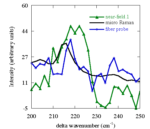
Normalized to peak at 767 cm-1. Both far-field measurements agree.
Surface Enhancement in NSOM-Raman Spectroscopy
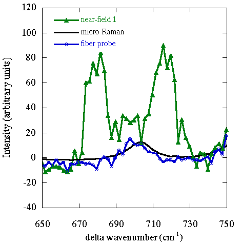
Normalized to peak at 767 cm-1. Both far-field measurements agree.
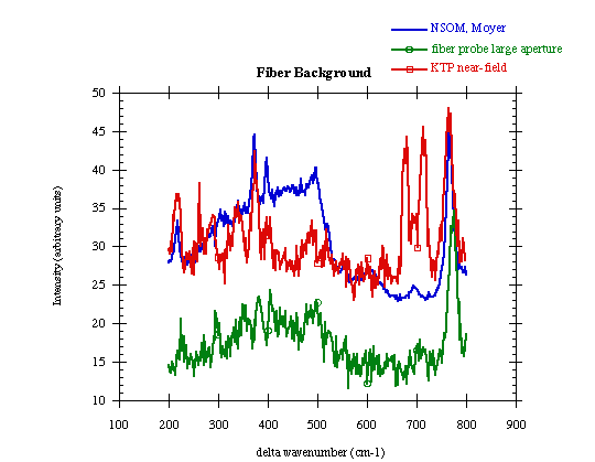
Background Raman Signal from the Probe
Relation to Probe Quality
Polarization
• The fiber output polarization is, in general, effected by the fiber. It depends upon the geometry of the tip, bends in the fiber, and the polarization of the light input to the fiber.
• A polarization-preserving fiber is not necessary (it has a very short length and the tip dominates).
• Use 'black-box' approach to set polarization -- measure output. Can obtain any probe output polarization in this way.
• E. Betzig et al. Appl. Opt. 31, 22 (1992).
• P.J. Moyer and M.A. Paesler, SPIE 1855, 58-66 (1993).
Higher sensitivity can be obtained by oscillating the polarization:
• M. Vaez-Iravani and R. Toledo-Crow, APL 63, 138-40 (1993).
• E.B. McDaniel, S.C. McClain, J.W.P. Hsu, Appl. Opt. 37, 84 (1998).
• D.A. Higgins et al. J. Phys. Chem. 100 (32) 13794-803
Polarization in the tip
• The metal coated tapered fiber probe tip supports a transverse electric mode.
• Some light polarized normal to the sample exists in the near-field region (theory verified).
• Any roughness on the coating will disrupt the tip polarization state or alter intensity vs. angle.
Metal samples with polarized light
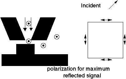
• Boundary conditions are the same as for the metal coating on the tip -- the tangential component of the electric field must be zero.
• This gives strong polarization effects.
• Transmission:
- for a long metal strip, light polarized parallel to the strip will tend to be reflected back up the fiber.
- light perpendicular to the strip will be able to couple around the edge and be collected.
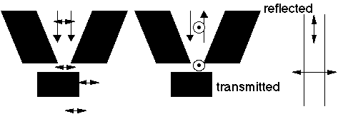
• Reflection - The new condition is that the light must make it way around the tip and up into the far-field.
- This changes the problem. Light levels are much smaller, and light polarized parallel to the strip are better able to couple out from under the tip and be reflected to the far-field.
• Reflection mode operation is much more susceptible to tip-induced artifacts than the transmission mode measurements.
- This is because the light must couple out from beneath the probe tip.
- One can get shadowing effects.
- Polarization data taken after a tip crash can show different behavior for the different polarization directions.
Dielectric samples with polarized light
• Less polarization dependence from small small structures.
• Birefringent samples: more.
Magnetic Imaging
• Light can be used to sense the magnetic field by use of the Faraday effect which rotates the polarization of the light.
• This can be done in either transmission or reflection.
• The polarization rotation can be small (~1°), but is observable (crossed polarizers without the sample).
• Image contrast can be reversed by rotating the analyzer (good check).
Early work -- magnetic bubbles and magneto-optic films
• E. Betzig, J.K. Trautman, J.S. Weiner, T.D. Harris and R. Wolfe, Applied Optics, 31, 4563-4568 (1992).
• E. Betzig, J.K. Trautman, R. Wolfe, E.M. Gyorgy and P.L. Finn, APL, 61, 142-144 (1992).
Recent magneto-optic imaging
• G. Eggers et al, Surf. and Inter. Anal. 75 (7-8) 483-7 (1997).
• C. Durkin, C. Lodder, I.V. Shvets, JAP 81 (8) 5019 (1997).
• M.R. Pufall, A. Berger and S. Schultz, JAP 81 (8) 5684-91 (1997).
Theory
• D. van Labeke, A. Vial and D. Barchiesi, SPIE 2782, 559-69 (1996).
A review of magnetic imaging with probe microscopes
• U. Hartmann, J. Magn. and Magn. Mater. 157-158, 545-9 (1996).
Data Storage
• A magnetic material is evaporated on a transparent substrate.
• The magnetic fields are all aligned in the same direction along the 'tracks'.
• The media is read by observing the rotation of the polarization of the NSOM signal.
• Bits are written by increasing the power input into the fiber probe.
• The tip of the probe heats up, and transfers energy to the sample locally. This raises the temperature of the magnetic film above the Curie temperature, and the direction of the field in the local region reverses (driven by the magnetic field from the neighboring areas).
• Read time is thus limited by the signal to noise for polarization detection (can be influenced by the topography).
• Write time is limited by the thermal transfer process.
• Seek time is limited by the speed of the feedback system to maintain the position of the probe in the near-field while avoiding a crash.
• Long term reliability is probably determined by the probe life, which is currently quite short even without the rapid raster rates.
• Density of bits is very high.
• To attain high enough rates, one may have to use several probes in parallel.
Biological Imaging
• NSOM holds much promise as many of the standard techniques developed for the far-field can be utilized by NSOM, including
- polarization contrast - index contrast - fluorescence (including all the dyes and methods for their attachment to cell structures). - luminescence - Raman spectroscopy - Infrared spectroscopy - Time-resolved measurements
• Fluorescence has been used to date.
• In addition, the topographic image provided by the force feedback provides additional information.
• NSOM is friendly in terms of imaging environment (water O.K.) and sample preparation (little required).
Early tissue slices, fibroblast cells.
• E. Betzig et al, Biophysics J. 49, 269-79 (1986).
• E. Betzig et al, Bioimaging 1 (3) 129-35 (1993).
• E. Betzig in Near Field Optics, D.W. Pohl and D. Courjon, eds. (Arc-et-Senans, France, Kluwer) 1993, 7.
Phospholipid monolayers
• Jeesong Hwang et al, Science 270 (5236) 610-44 (1995).
'Intact' myofibrils
• E.J. Seibel and G.H. Pollack, J. Microsc. 186 (3) 221-31 (1997).
Chromosomes
• M.H.P. Moers, W.H.J.Kalle, N.F. Van Hulst, J. Micr. 182 p1,40 (1996)
• N.VanHulst,M.Garcia-Parajo,A.Ruiter,J.Struct Bio.119(2)222(1997).
• W. Wiegrabe, et al. Surf. and Interf. Anal. 25 (7-8) 510-13 (1997).
In addition, much work has been done on organic films and
• Limitations for biological imaging:
- NSOM is a surface sensitive technique. Cell membranes Thin parts of cells such as moving cells (fibroblast cells work well).
- Cells can move. This is equivalent to microscope drift. Requires rapid imaging and a fast feedback loop.
Problem: many of the high power techniques require slow imaging to obtain adequate signal to noise.
Solution options: drug or kill the cells to slow them down, live with low signal to noise images, work with larger probes and sacrifice resolution.
- The effect of a possibly hot tip nearby has not been completely considered.
- Resolution may not be high enough for use on single molecules in an imaging sense. (although single molecules can be detected)
Quantum Dots, Quantum Lines
• Quantum structures are usually small and studied at low temperatures.
• NSOM has adequate resolution, and instruments have been cooled.
• Two modes are possible, illumination (which can be plagued by carrier diffusion) and collection of the structure luminescence.
Some of the first low temperature instruments:
• R. D. Grober et al, APL 64 (11) 1421-3 (1994).
• H. Ghaemi, C. Cates, B.B. Goldberg, Ultramicr. 57, 165-8 (1995).
Spatial & energy & magnetic field resolved states in a single quantum dot:
• H.F. Hess et al, Science 264 (5166) 1740-5 (1994).
Individual quantum structures
• H.F. Ghaemi et al, Superlattices and Microstr. 17 (1) 15-16 (1995)
Single wires
• T.D. Harris et al, APL 68 (7) 988-90 (1996).
• T.D. Harris et al, Semic. Sci. and Techn. 11 (115) 1569-74 (1996).
Recent dot theory
• B. Hanewinkel et al, Phys Rev B55 (20) 13715-25 (1997).
Recent cleaved edge overgrowth wires
• M. Katz, et al, J. Luminescence 72-74, 12-17 (1997).
Recent spectroscopy on wires
• A. Richter et al, Surf. and Interf. Anal, 25 (7-8) 583-92 (1997).
Lithography
• The spatial resolution makes NSOM a natural choice for high resolution lithography.
• Caveats are similar to data storage:
- Write time is limited by the small signal and by the speed of the feedback system to maintain the position of the probe in the near-field while avoiding a crash.
- Long term reliability is probably determined by the probe life with rapid raster rates.
- To attain high enough rates, one may have to use several probes in parallel.
• Another active issue is the choice of resist
- It cannot be thick, must be active in the range of wavelengths the probe emits, et cet.
Some references:
• S. Wegscheider et al, Thin Solid Films 264 (2) 264-7 (1995).
• S. Davy, G Rach and H. Spajer SPIE 2782, 551-8 (1996).
Direct write oxidation of H-terminated Si
• S. Madsen et al, APL 69 (4) 544-6 (1996).
• S. Madsen et al. JAP 82 (1) 49-53 (1997).
Oxidation of polymers
• P.K. Wei, et al. Adv. Mat. 8 (7) 573-6 (1996).
Conjugate and electroluminescent polymers
• E.Z. Faraggi et al, Synth Metals 85 (1-3) 1187-90 (1997).
• P.K. Wei et al, Synth. Metals 85 (1-3) 1421-2 (1997).
Photonics Device Characterization
• To reduce dissipation in electro-optic devices, one wishes to reduce the threshold current, so builds very small lasers.
• Studying these lasers is difficult if only the far- field data is used.
• NSOM can bridge the gap between the lithography and the far-field radiation pattern by allowing one to study the light intensity profile in the near-field and transition regions.
• Dopant profiles can be investigated by observing the luminescence and lasing in the near field -- the functional forms of the intensity decay are different for luminescence and lasing
• Wave guide leakage can also be explored.
Early laser image, including a hot spot seen with topography
• S.K. Burratto et al. JAP 76, 7720 (1994) & APL 65, 2654 (1994).
Waveguides
• A.G. Choo et al, Ultramicroscopy 57, 124-9 (1995).
Aging of lasers
• A Richter, Ch. Lienan, J.W. Tomm, Surf & Interf. Anal. 25 (7-8) 573-82 (1997).
E-fields
• J.K. Leung, C.C. William and J.M. Olson, Phys Rev B56 (3) 1472-8 (1997).
Lasers and luminescence
• Lin Jutmy et al, APL 69 (23) 3519-21 (1996).
• I Fischer et al. Europhys Lett. 35 (8) 579-84 (1996).
• M. Ohtsu et al, JJAP 36 (7B) pt 2 L896-8 (1997).
Semiconductors/ Defect Characterization
• NSOM has been used to extend several characterization techniques to higher resolution, here is a sampling:
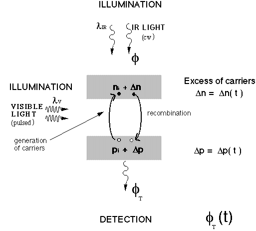
All-optical carrier lifetime measurements
• A. LaRosa, B. I. Yakobson, H.D. Hallen, APL 70 (13), 1656 (1997).
• A. LaRosa, C.L. Jahncke, and H.D. Hallen, SPIE 2384, 101 (1995).
• A. LaRosa, C.L. Jahncke, and H.D. Hallen, Ultramicroscopy 57, 303-308 (1995).
NOBIC (Near-field optical beam induced carriers)
• Xu Qin, M.H. Gray, J.W.P. Hsu, JAP 82 (2) 748-55 (1997).
Threading dislocations with polarization studies
• J.W.P. Hsu et al, APL 65 (3) 344-6 (1994); JAP 79(10) 7743 (1996).
Solar cells
• A.A. McDaniel, J.W.P. Hsu, A.M. Gabor, APL 70 (26) 3555-7 (1997).
SOI (silicon on insulator)
• A. LaRosa, B. I. Yakobson, H.D. Hallen, Proc of Diagn Tech for Semic Mater Proc II (Boston, MA, 27-30 Nov 1995) Mat. Res. Soc. Symp. Proc. 406, 189-194 (1996).
Spectroscopy
• W.M. Duncan, above MRS meeting, 183-8.
• B.B. Goldberg et al, above MRS meeting, 171-82.
• H.D. Hallen, A. LaRosa, C.L. Jahncke, Phys Stat Solidi (a) 152, 257-268 (1995).
• C. Coluzza et al, SPIE 2782, 591-601 (1996).
Surface passivation
• J. Liu and T. Kuech, APL 69 (5) 662-4 (1996).
• J. Liu and T. Kuech, above MRS meeting, 523-8.
Devices
• R.M. Cramer et al, Proc. 22nd Int. Symp for Testing and Failure Anal, 19-24 (Los Angeles, CA, 18-22 Nov 1996).
• J.P. Fillard, DRIP95, p. 195-200 (Boulder, CO, 3-6 Dec 1995).
Time Contrast
Optical Measurements of Recombination Time Constants
• A. LaRosa, B. I. Yakobson, H.D. Hallen, APL 70 (13), 1656 (1997).
• Visible light creates carriers, infrared light monitors.
Optical Measurements of Recombination Time Constants
-- The Experimental Set-Up --
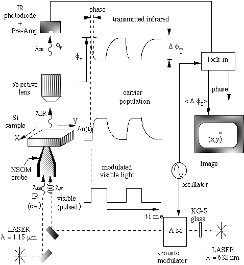
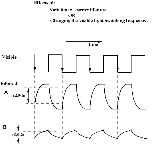
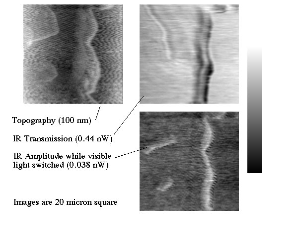
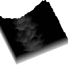
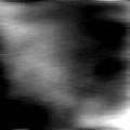
The time constant (white faster, lower right) reflects the topography (upper left) and is different than the (less than a percent variation) IR NSOM image (upper right).
Variation of the Visible Light Chopping Frequency
• The lifetime can be quantitatively measured from
the 'knee' position (2 samples shown).
• Images at different frequencies highlight the slower
lifetime process or the faster diffusion process.
Shown are 7.5
mm square images at 100, 2k, 20kHz.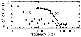

• The upper right image is
l-square (1.15 micron).Silicon-on -Insulator
• A. LaRosa, B. I. Yakobson, H.D. Hallen, Mat. Res. Soc. Symp. Proc. 406, 189-194 (1996).
• Some regions reflect carrier lifetime as above.
• Other regions, of less uniform thickness, also reflect
the ability of the carriers to diffuse away -- thickness
variations:
(a) (b) (c)
(d) (e) (f)
All images correspond to the same region of 7.5
Spatial Resolution in Carrier Lifetime Studies
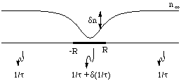
• Question:
- Why do we obtain high resolution when carrier diffusion is so large?
- With D = 10 cm
• Model:
- The carriers, once formed locally by the visible light, rapidly diffuse.
- The diffusion profile reflects the local time constant.
- Estimate the contrast
- Or consider a simple case:
t=0 inside a sphere of radius R, t=• outside. The profile n/n0 ~ 1 - R/r depends on R not D.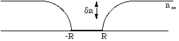
- D is reflected in the contrast.
![]()
North Carolina State University | Physics | Optics Home
Last updated on September 27, 2000