![]()
![]()
The Optics Laboratory
Group of
Hans Hallen, North Carolina State University Physics Department![]()
Surface Enhancement in Near-Field Raman Spectroscopy
The NSOM is a good tool for studying surface enhancement, since the probe can be moved toward and away from the surface with very high precision. Force feedback can be used to stabilize the position if the probe is close enough to the surface. We expect that Raman spectra, and most other optical signals, will be enhanced as the NSOM probe approaches the surface, since there are many evanescent modes of the electric field near the tip than can begin to interact with the sample.
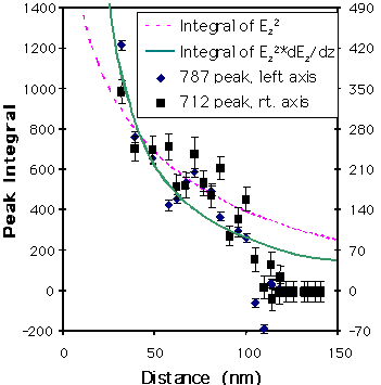
A good starting point is the Bethe-Bouwkamp model for electric fields near an aperture. To calculate the distance dependence, we use this model to calculate the electric field above the surface, then average the relevant quantity for all points in each plane above the surface, giving us one averaged value per distance,
 .
.
Independent calculation of the components is meaningful, since the vibration is usually along a bond, and only one component of the field will couple to the bond when the bond happens to line up with that axis. The squares of the projected fields appear in the Raman intensity, so they are shown. The calculation assumes light polarized along the x-direction incident on an aperture in the xy-plane. The plot with the derivative of Ez is what is expected in gradient field Raman, for comparison.
Below is a nano-Raman spectrum taken with our cooled CCD apparatus.
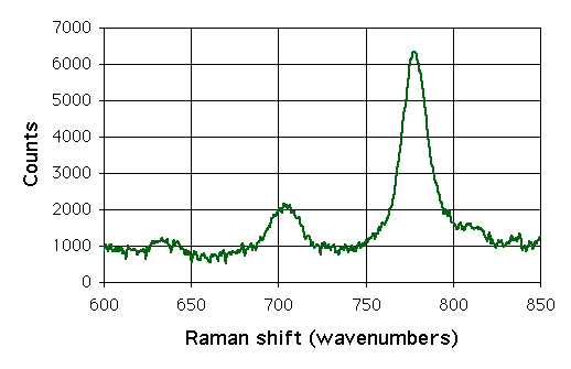
We take spectra similar to this at several distances from the surface, and subtract those taken closer from the average of several taken with the probe retracted from the surface – in the far-field. The difference spectra below represent the change as the probe enters the near field.
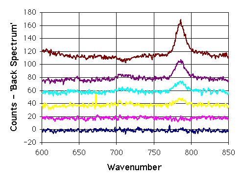
The spectra are shifted by 20 counts between each, and the spectra closer to the surface are towards the top of the figure. There are enhancements as one approaches the surface. At first one might think that these are simply enhancements of the existing Raman lines. They are not, since they are at different energies. Nor are they shifted by the same amount from the existing lines. They are at the energies of vibrations in KTP that are different than the vibrations observed in the far-field spectra. Two questions arise:
![]() Why aren’t the far-field Raman peaks enhanced? The top figure shows enhancement in all the electric fields, including X and Y, which are responsible for far-field Raman.
Why aren’t the far-field Raman peaks enhanced? The top figure shows enhancement in all the electric fields, including X and Y, which are responsible for far-field Raman.
![]() Where do these new peaks come from?
Where do these new peaks come from?
 ,
, .
. ,
, .
. ,
,