![]()
![]()
The Optics Laboratory
Group of
Hans Hallen, North Carolina State University Physics Department![]()
Nano-Raman Imaging
We image with near-field Raman in a manner very similar to typical near-field scanning optical microscopy NSOM imaging, except the output light is directed to a spectrometer before being analyzed. This is the apparatus used for this work:

The NSOM collects light in both transmission and reflection. The transmitted light is focussed into a photomultiplier tube (PMT) to provide a typical NSOM reference image. Contrast in this image will be related to the optical properties of the sample, refractive index and absorption variations. The reflected light is collected in an efficient, symmetric manner by drilling a hole through a 0.55 NA aspheric lens, and gluing in the tip. (Actually, we superglue to a hypodermic tube, which is epoxied into the glass.) An image is formed serially by scanning the tip over the sample. Topography is measured by using force feedback to adjust the tip-sample separation to a constant value, and recording the motion required. This feedback is necessary to keep the tip in the near-field of the sample. The sample is potassium titanyl phosphate (KTP) that has been Rb-doped in squares 5 microns on a side and spaced by 5 microns. The doping induces stress in the material, giving a 12 nm uplift in the Rb-doped regions that we can see in the following 12 micron square topographic image:
 .
.
The image is color-coded to black-red-orange-yellow-white with the yellow ~12 nm higher than the red. We used this image to locate the tip for taking Raman spectra both on, and off, the Rb-doped region:
 .
.
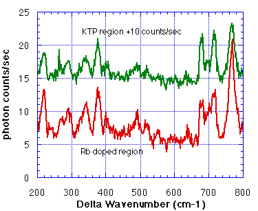
The spectra are quite similar, although a clear difference exists in the height of the peak at 767 wavenumbers relative to the other peaks. We therefore set the spectrometer to record this wavelength, and began scanning. Three images were simultaneously collected: the topography on the left, indicating the corner of one of the Rb-doped regions, the NSOM transmission image in the center, indicating the index of refraction variations, and next the first nano-Raman image ever taken. The Raman image was taken with reflected light, and reflects the different chemical bonding in the Rb-doped region. The images are 4 micron square.

The few percent change (we subtracted a large flat background) of the NSOM-transmission image shows the utility of the Rb-doping. It changes the index of refraction, and is used to form light-guides on the KTP surface. The few percent higher index of refraction of the RTP in the KTP serves to pull more light from the NSOM tip, and also to hold light for lateral propagation along a channel. The Raman image contrast is what would be expected from the individual Raman spectra: the signal is higher over the Rb-doped regions.
Due to the low signal levels (recall Raman is ~10^(-6) and NSOM intensities are 1-100 nW), we had to integrate the photon counts for 20 seconds at each image point. This means that taking this set of images was a ~10 hour job. One worries about our ability to reduce thermal and other motions of the tip so that the image is not smeared during this time. It is a difficult task to do so, but we stabilized the instrument so that it drifts less than 200 nm in 10 hours, and verified that in fast topographic images before and after each long image. The other detail that you might notice in the images is a shift between the topographic and optical images. This is due to that fact that the probe is never exactly perpendicular to the surface, so the topography s recorded off a corner of the probe tip, whereas the optical image is referenced to the center of the probe tip.
![]()
NC State University | Physics | Optics Home
Last updated on September 29, 2000
Copyright ©2000, Hallen Laboratory, NCSU, Raleigh, NC. www.physics.ncsu.edu/optics
Comments or questions? Hans_Hallen@ncsu.edu