![]()
![]()
The Optics Laboratory
Group of
Hans Hallen, North Carolina State University Physics Department![]()
Near-field vs. Far-field
Raman Spectroscopy
Here we catalogue the
differences between near-field and far-field Raman spectroscopy. They are:![]()
![]()
![]()
![]()
![]()
![]()
![]()
To compare the Raman spectra, we studied potassium titanyl phosphate (KTP) under three conditions: micro-Raman (far-field), NSOM with the tip pulled away from the sample (far-field), and NSOM with the tip in the proximity of the sample (near-field). The far-field NSOM case was necessary to insure that the fiber or other aspects of the measurement system were not effecting the outcome. It should resemble the other far-field measurement. The spectra --

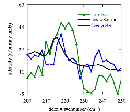
One can see differences in these spectra. In particular, one can see the Rayleigh tail on the left of the micro-Raman spectra (black), and a noticeable difference in the near-field spectrum (green) near 700 wavenumbers, but one cannot ascertain whether scaling accounts for some of the differences, and whether the two far-field measurements are the same. We therefore scale the spectra so that they agree on the big peak at 767 wavenumbers, and look at the parts of these spectra. We first examine the region of the lower energy vibrations:

The two far-field spectra agree to within noise, as expected. The near-field spectrum has a lower background, more area under the peak, and a shift to higher energies of the peak. All these changes are significant. The lower background results from the smaller, hence more uniform, region sampled in the near field. With less inhomogeneities, there is less Rayleigh scattering and hence less Rayleigh tail. The other differences result from the same effect -- a change in selection rules as a result of the different nature of light in the near-field. The metal aperture on the NSOM alters the direction of the electric field since the electric field must be normal to the metal surface, not tangential. At the very tip, this corresponds to a longitudinal, or Z component, of the electric field, in contrast to the transverse propagating fields. The z-component couples to a vibration mode at 220 wavenumbers, one that is not coupled to by the transverse field (that couples to the mode at 215 wavenumbers present in the far-field spectra). This additional mode increases the area under the peak, and since the addition is at higher energy, shifts the peak.
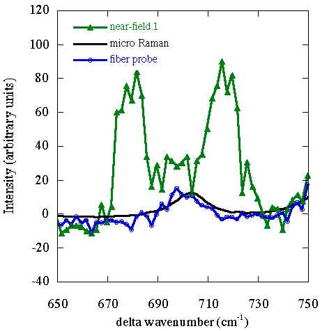
The situation at higher photon energy shifts, at higher vibration energies, is somewhat different:

The backgrounds are all the same, since the Rayleigh tail has diminished. The two far-field measurements agree (good). The near-field spectrum contains two peaks that are not in the far-field spectra. Arguably, the far-field peak is still present, although overshadowed by the new peaks. The new peaks cannot be simply explained as those at lower energies, since no Raman peaks of sufficient strength have been observed with electric field in any direction. We have derived a model that describes the origin and strength of these peaks originating from the Z-component electric field gradient near the NSOM tip. We call it gradient field Raman, and describe it on an accompanying web page.
![]()
![]()
Last updated on September 29, 2000
Copyright ©2000, Hallen Laboratory, NCSU, Raleigh, NC. www.physics.ncsu.edu/optics
Comments or questions? Hans_Hallen@ncsu.edu