![]()
![]()
![]()
![]()
The Optics Laboratory
Group of
Hans Hallen, North Carolina State University Physics Department![]()
NSOM Electromigration in YBCO
Our method for electromigration with the NSOM and for data processing have been discussed in the preceding web pages.
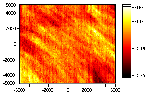
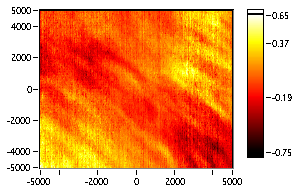
In one case, after applying -1 volt to the sample with a tunnel current of 2 nA for 36 minutes, we observed the following electromigration:
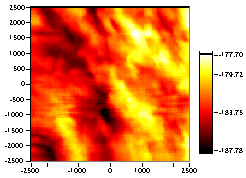
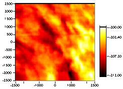
The left image is the optical image before electromigration, and the image on the right is afterwards. The x&y scales are in nanometers, and the vertical (oxygen) scale in arbitrary units. We shift the images, scale, and subtract to view the region effected by the EM:
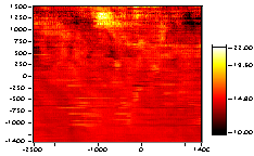
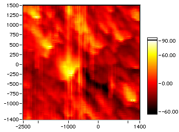
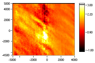
The left image shows the increase in oxygen under where the tip had been, and the right image the corresponding topography in nm for the overlapping region of the two images above.
This analysis facilitates measurement of the 240 nm radius of the electromigrated region.Another electromigration used a much higher dose. After applying -3 volt to the sample with a tunnel current of 3 nA for 8 hours:
The left image is the optical image before electromigration, and the image on the right is afterwards. The x&y scales are in nanometers, and the vertical (oxygen) scale in arbitrary units. We shift the images, scale, and subtract to view the region effected by the EM:
Again, the bright spot at the center shows a 250 nm radius region in which the electromigration occurred. Comparison with the topography in both cases shows that the EM is limited in size to one grain.
We can learn a little more about the granular dependence of electromigration by watching the motion of oxygen contour lines when they are plotted on top of a color scale of the topography:
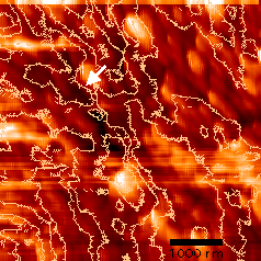
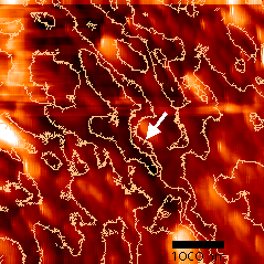
The two white arrows point to the same grain, the left is before electromigration and the right after. Note the contour lines to the right of the grain have been pulled into the grain, that is, the grain’s oxygen concentration has increased. The tick marks on the contours point towards lower oxygen concentration.
The following information about the mechanism for oxygen motion can be learned from the above data:
"electron wind"
"Electric field"
"hot" electrons
Possible size limitations:
The ‘hot electron’ mechanism describes all the data. It is interesting to note that a similar mechanism was found for hot electron induced motion of gold atoms.
![]()
![]()
Last updated on October 5, 2000