![]()
![]()
The Optics Laboratory
Group of
Hans Hallen, North Carolina State University Physics Department![]()
GFR in NSOM-Raman
Near-field scanning optical microscopy (NSOM), using a metal aperture at the probe tip to limit the illuminated area, provides an experimental configuration where GFR effects, including those of solid materials rather than molecules, may be observed. The metal that forms the aperture creates the strong field gradients that are required. This configuration has the advantage that the metal can be moved with high precision in all three dimensions. It can be scanned over a surface, permitting studies of adsorbed species or the solid substrate itself. Further, it can be retracted from the surface so as to move the high field gradient region away from the surface, effectively ‘turning off’ the GFR effect. This is an important test to confirm the origins of the observed Raman lines.
In NSOM, a sharpened optical fiber is coated with metal by rotating the probe with the tip angled away from the evaporation source so that an aperture is left uncoated. We have used fibers sharpened by the heat-and-pull method and by etching. Aluminum forms the aperture. The probe is positioned near the surface under lateral force feedback. The NSOM is used in illumination mode, with 514 nm Ar ion laser light. Reflected light is collimated with a 0.50 NA lens and focussed into a 1 meter, Czerny-Turner spectrometer. The primary difficulty encountered in NSOM-Raman is that of low signal levels. Input of more than a few milliwatts of light into the probe’s fiber will destroy the probe tip. Smaller tip apertures strongly reduce the probe throughput, and Raman cross sections are relatively small. The aperture size directly controls the lateral resolution of the microscope.
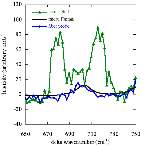
We have taken comparative spectra of a KTP sample using micro-Raman, Raman with an NSOM probe retracted from the surface, and NSOM-Raman (in the near-field). Portions of these spectra are

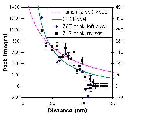
The spectra have been normalized to the strong Raman peak at 767 cm-1. The two far field spectra are the same to within noise, but the near-field spectrum differs by the apparent addition of peaks near 680 and 714 cm-1. KTP has vibration energies at 683 and 712 cm-1. The 683 line has been observed as a weak line in prior Raman work. The 712 wavenumber vibration has been observed as a strong IR (not Raman) line. The distance dependence (from a later study),

is consistent with the field gradient model. The Bethe-Bouwkamp model for NSOM fields is used to derive the expected GFR and standard Raman distance dependence in the figure. The GFR model provides a much better fit. Also, since the 712 cm-1 mode is strong in IR absorption, it is likely that the GFR effect is at least partially responsible for the spectra. The ratio of the Raman peak at 767 cm-1 to the GFR peak at 712 cm-1 is about 2 for the data in the first figure, and ~200 for that in the second figure. These ratios compare well to those of a simple microscopic model based upon polarizabilities from the literature, and polarizability gradients estimated from comparison of the long and short Ti-O bond data. The model gives a ratio of ~2.8 when the field gradient / field ratio is calculated for a flake on the tip (an edge 1Å from the surface), and ~170 for the Bethe-Bouwkamp fields at 10 nm.
More info is in the papers.![]()
Last updated on October 2, 2000
Copyright ©2000, Hallen Laboratory, NCSU, Raleigh, NC. www.physics.ncsu.edu/optics
Comments or questions? Hans_Hallen@ncsu.edu